A biosensor is mainly composed of three elements:
i) the biological recognition element (enzyme, antibody, DNA, micro-organism);
ii) the transduction element relying on electrochemical, optical, piezoelectrical or thermal principles;
iii) the signal processor.
The specificity of a biosensor depends exclusively on the bioreceptor molecule, while the sensitivity, stability and reliability are mainly determined by both, the nature of the bioreceptors and the type of the physical transduction system.
We use different surface chemistry protocols to immobilize the bioreceptors (i.e. antibodies, DNA or aptamers) on the sensor surface in order to achieve specific, reproducible and accurate biosensors. For example to functionalize gold substrates with antibodies we normally use self-assembling monolayers (SAM) followed by EDC/NHS chemistry or streptavidin-biotin binding (Figure 2).
Figure 2: A schematic diagram showing the functionalization of a gold substrate with antibodies for the detection of a certain target protein.

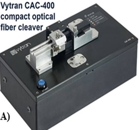
The Vytran CAC-400 compact fiber cleaver (Figure A) is designed for cleaving optical fibers (FO) with claddings from Ø60 μm to Ø600 μm, with a high degree of accuracy, ease of use and versatility. CAC400 device produces flat cleaves perpendicular to the length of the FO. The unit operates with a tension-and-scribe cleave method, whereby axial tension is first applied to the FO followed by an automated scribe process utilizing a diamond cleave blade. After the blade scribes the FO, the tension is maintained, causing the scribe to propagate across the FO perimeter and complete the cleavage.

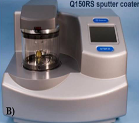
The Q150RS rotary-pumped sputter (Figure B) coater is fully automated, compact and suitable for Scanning Electron Microscopy (SEM) and other applications requiring thin films of non-oxidising metals (i.e. gold, silver, platinum and palladium).
For a higher level of control and reproducibility, the film thickness monitoring option allows the user to pre-determine de coating thickness (in nanometres).